

Structure of Metaphasic Chromosome
During the prophase and metaphase stage of cell- division the chromatin undergoes process of compactation and condensation in order to deliver discrete packets of chromatin to daughter cells.
During the process of compactation chromatin acquire predictable and visible structure depending upon the length of DNA.
Metaphasic chromosome is highly condensed chromatin consisting of two molecules of condensed chromatin called sister chromatids.
It is highly organized and reproducible structure with distinct morphology that can be seen under compound microscope.
The metaphasic chromosome can be differentiated into two chromatids each containing identical copy of double stranded DNA associated with histone and non-histone protein.
The two sister chromatids are held together at a specific point called centromere which is represented by primary constriction.
Centromere
- Centromere is the region of metaphasic chromosome where two sister chromatids are held together. It also provides the binding site for the spindle fibres during the cell division.
- Centromere is heterochromatic region containing unique chromatin consist of DNA associated with H3 histone variant called CENP-H3.
- The centromeric DNA codes for specific proteins that forms kinetochores where spindle fibres are attached.
- The kinetochores acts as motor proteins that drags the two sister chromatids towards the opposite poles along the spindle fibres.
- Majority of chromosome contain single centromere and they are called monocentric chromosome.
- In some organisms like Ascaris, Junacales, Cyperales the centromeric activity is diffused throughout the surface of chromosome and such a chromosome is called Holocentric or Holokinetic chromosome.
Chromosomes can be classified on the basis of position of centromere and accordingly chromosomes are of four types:
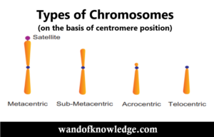
- Metacentric chromosome: It has two identical arms and centromere is present at the centre of two arms. It is V-shaped.
- Sub-metacentric chromosome: Its two arms are of unequal length and centromere is present at sub-centric position. They are J or L shaped.
- Acrocentric: Centromere is sub-terminal in position. It is I- shaped.
- Telocentric: Centromere is terminal in position. It is also I-shaped.
Telomere
- The terminal end of eukaryotic chromosome is called telomere.
- The telomere maintains the structure and replication of the DNA of each chromosome.
- It prevents the fusion of the DNA of different chromosomes.
- The terminal end of the eukaryotic DNA contains tendum repeats of G-nucleotide rich region called telomeric DNA. This DNA codes for enzymes telomerase which is a reverse transcriptase enzyme that extends the 3’ end of DNA by providing RNA template.
- The extended 3’ end acts as template for the extension of complementary strand which become shortens after the removal of primer. It is called solution of end replication problem.
- The extended 3’ end folded on itself forming a completely closed loop that protects the terminal end of the chromosome. This loop in association with protein securing forms a cap like structure at the terminal end of chromosome called telomere.
Important links
- Molecular Structure of Eukaryotic Chromatin| Levels of DNA packaging
- Succession of plants in Water or Hydrosere or Hydrarch
- Digestive System of Balanoglossus (Hemichordata)
- Scales of Fishes | Modification and Uses of Scales
- Habit and Habitat of Rana tigrina (Indian Bull Frog)
- Affinities of Hemichordata with Chordata| Annelida & Echinodermata
- Reproduction in Protozoa- Asexual & Sexual Reproduction
- Parasitism in Protozoa |Types of Parasites |Host specificity
- Bioremediation: Meaning | Need | Merits | Scope & Approaches
- Landform: Types| Processes in Formation| Gradation of Landform
- Radioactive Substances |Units of Radioactivity & Its Effect
- Environmental Biology-Scope & Major Areas of Environmental Biology
- Classification of Invertebrates & General Characteristics
Disclaimer: wandofknowledge.com is created only for the purpose of education and knowledge. For any queries, disclaimer is requested to kindly contact us. We assure you we will do our best. We do not support piracy. If in any way it violates the law or there is any problem, please mail us on wandofknowledge539@gmail.com